I have realized that there are a couple of errors in my book “Athletic and Working Dog: Functional Anatomy and Biomechanics”. I would like to provide the corrections for these errors. I apologize for this and am grateful to the readers who have recognized them and brought them to my attention.
On page 49, the paragraph beginning at the bottom of the left column is incorrect, the last sentence incorrectly lists muscles that are not located in the rear limb:
“Many of the muscles associated with actions of the distal extremity originate in the region of the stifle (Figure 59). The muscle that is typically the largest of these muscles is the gastrocnemius. The lateral head originates at the large tendon of the lateral supracondylar tuberosity of the femur. The medial head arises on the medial supracondylar tuberosity of the femur. The muscle distally converges into the calcaneal tendon which inserts at the tuber calcanei. It primarily acts to extend the tarsal joint and slightly flex the stifle joint. [The other antebrachial muscle that originate in this area are the: Tibialis Cranialis, Peroneus Longus, Peroneus Brevis, Extensor Carpi Radialis, Extensor Digitorum Communis, Extensor Digitorum Lateralis, Extensor Digitorum Longus, Ulnaris Lateralis, Tibialis Caudalis, Flexor Hallucis Longus, Flexor Digitorum Profundus, Medial Head and Lateral Head, Flexor Digitorum Superficialis and the Flexor Carpi Radialis.]”
The last sentence should read:
“The other antebrachial muscles that originate in this area are the: Tibialis Cranialis, Peroneus Longus, Peroneus Brevis, Extensor Digitorum Lateralis, Extensor Digitorum Longus, Extensor Digitorum Hallucis Longus, Flexor Hallucis Longus, Flexor Digitorum Profundus, Flexor Digitorum Superficialis.”

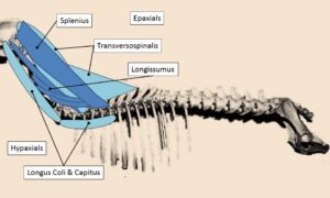
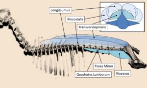
On page 54-55, Figures 64 & 65 “the axial muscles of the cervicothoracic region” correctly shows the Hypaxial and Epaxial muscle groups. Below the figure the text in the narrative below is incorrect and has it swapped.


The text below it should read:
In the cervicothoracic region the “epaxial” muscles include the transversospinalis, splenius and the longissimus muscles (Figure 64). The “hypaxial” muscles of the cervicothoracic region include the longus coli and capitis. In the thoracolumbar region (Figure 65) the “epaxial” muscles include the transversospinalis, iliocostalis and the longissimus muscle groups. The “hypaxial” muscles of the thoracolumbar region are the quadratus lumborum, psoas minor and the iliopsoas.
